
Doctors are quick to recognise osteoporosis in women, but often overlook it in men.
A 67-year-old man in your practice is referred back to you for follow-up after a distal radius fracture.
What is the key information you should elicit from him, and what is the key information you should convey to him?
The patient in this scenario is at elevated risk for another fracture and, according to some, for death. (1-3)
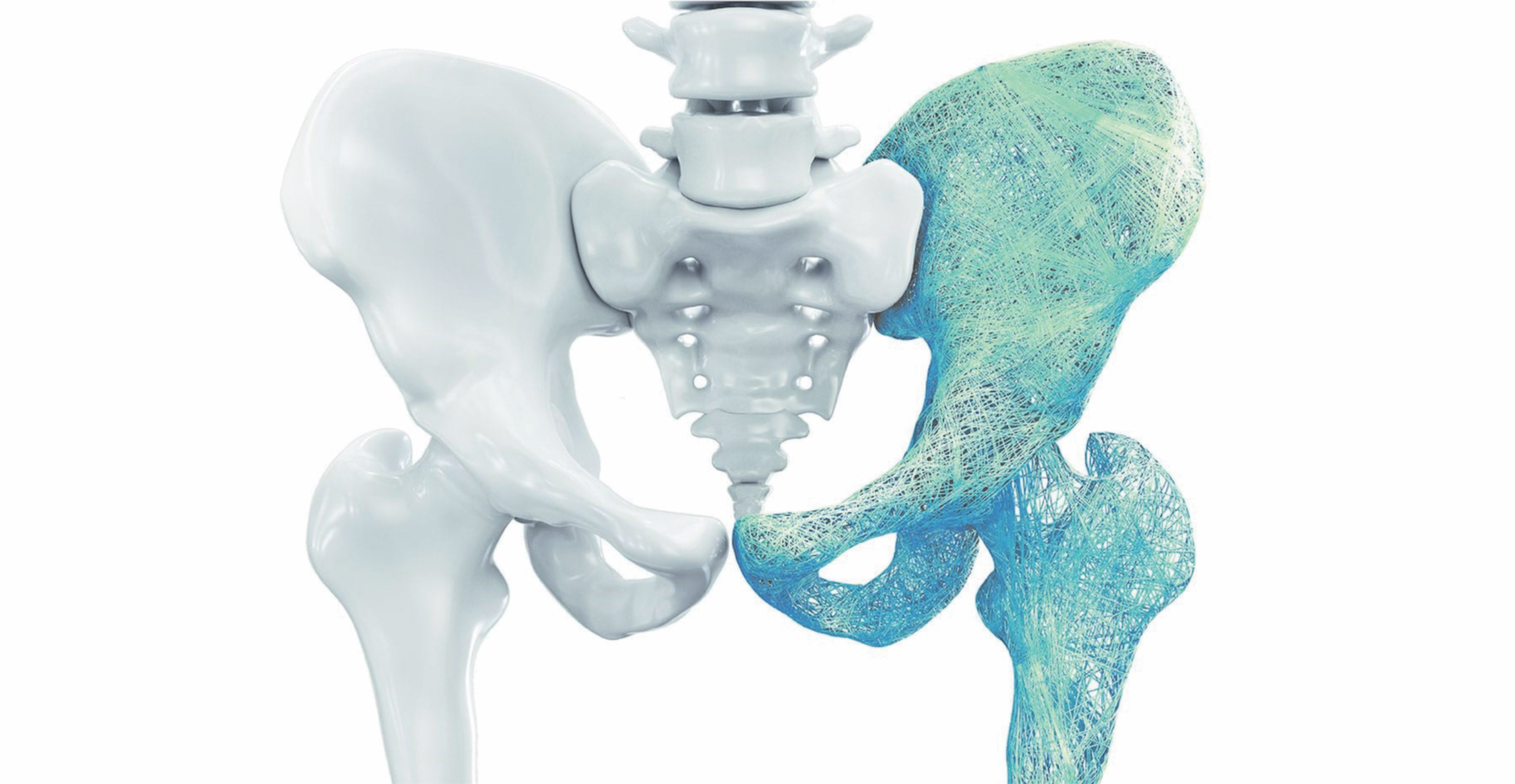
Fractures are common and prior fracture is one of the most powerful risk factors for subsequent fracture. A fracture in an adult patient, particularly over the age of 50 years, should always prompt investigation.
There are several widely used fracture risk calculators, with FRAX and the Garvan calculator being most popular in Australia. These decision support tools can be used without a prior measurement of bone mineral density (BMD) and take only seconds to complete at the point of care.
Neither accounts for the most recent conceptual advance in management: imminent fracture risk.
Fracture patients are at the highest risk of subsequent fracture soon after the index fracture, and this has led to special emphasis on secondary fracture prevention, as operationalised by fracture liaison services and similar initiatives. (4, 5)
So, what about our patient?
The first thing is to learn the details of how the fracture was sustained. In the past, much attention was given over to determining whether the fracture was a “fragility” or “low-trauma” fracture, these synonymous terms being defined by whether the trauma was a fall from standing height or less. A fracture under these circumstances should always be considered abnormal, regardless of age and the circumstances leading to the fall.
Most patients will blame the fracture on the trauma, saying something like, “Anyone would have fractured if they’d fallen the way I did.” That is nearly always incorrect, and your first job is to communicate that the fracture is a manifestation of underlying osteoporosis unless another, identifiable secondary cause of skeletal fragility is present.
This is analogous to atherosclerotic disease providing the substrate for cardiovascular and cerebrovascular events, even though the underlying disease had previously been clinically silent.
Recent data calls into question whether the “fragility” and “low-trauma” qualifiers are as meaningful as previously believed, as even high trauma fractures have been shown to increase subsequent fracture risk. (6)
A complete personal and family history of fractures should be obtained, as should a falls history. Comorbidities, medications, diet, exercise, and habits should be reviewed, with counselling regarding dietary calcium and protein consumption, vitamin D, smoking, and drinking provided. Height loss, and congruence of stated and measured height should be assessed. Strength, balance, and ambulation should be assessed.
Ancillary testing should include bone densitometry if not obtained within the prior year, blood chemistry, blood cell counts, 25 hydroxyvitamin D level, and 24-hour urine creatinine and calcium.
If there are existing radiographs, CT scans, or MRI scans that allow visualisation of the vertebral column, these images (not the reports) should be reviewed to seek prevalent vertebral fractures. Only about 1/3 of vertebral fractures come to clinical attention at the time of their occurrence and many are not reported on imaging. (7)
If there has been historical height loss of 4cm or incident height loss of 2cm, new vertebral imaging should be obtained. Because the risk of recurrent fracture is greatest immediately following the index fracture, treatment should be initiated promptly, beginning even before bone density has been measured.

Where can you find more information?
The Asia-Pacific Fragility Fracture Alliance (APFFA) is a federation whose members include a number of regional professional societies, the worldwide Fragility Fracture Network, the International Osteoporosis Foundation, and the International Society for Clinical Densitometry. Together, the member organisations have committed to working collaboratively to reduce the burden of disease caused by fractures in the region.
To this end, we have developed an educational toolkit for primary care physicians. The toolkit provides everything needed to present a half day educational program geared toward general practitioners. Included are slides for two 60-minute lectures, the first on the burden of disease caused by fracture and the second on the evaluation and management of patients who have suffered recent fractures.
There are materials for leading discussions that address local variations in resource availability, and templates for meeting invitations and completion certificates. All of these are available at the APFFA website (https://apfracturealliance.org).
APFFA has other ongoing projects, including development of a regional hip fracture registry and research projects aimed at precisely defining the burden and patterns of management following hip fracture in the Asia-Pacific region. APFFA is hosting a more comprehensive collection of educational materials as well, addressing specific topics of interest to non-GP specialist physicians as well as general practitioners. These are primarily materials that have been developed by others, links to which are hosted by APFFA.
This approach is laid out in greater detail in the toolkit and the references cited here.
In addition, I have covered the primary care approach to fractures and osteoporosis in peri- and postmenopausal women. (8)
Rob Blank is Professor Emeritus of Medicine (Endocrinology), Physiology, and Cell Biology at the Medical College of Wisconsin, Visiting Scientist at the Garvan Institute of Medical Research, and past president of the International Society for Clinical Densitometry.
References:
- Morin S, Lix LM, Azimaee M, Metge C, Caetano P, Leslie WD. Mortality rates after incident non-traumatic fractures in older men and women. Osteoporos Int. 2011;22(9):2439-48.
- Kanis JA, Johansson H, Oden A, Harvey NC, Gudnason V, Sanders KM, et al. Characteristics of recurrent fractures. Osteoporos Int. 2018;29(8):1747-57.
- Hansen L, Petersen KD, Eriksen SA, Langdahl BL, Eiken PA, Brixen K, et al. Subsequent fracture rates in a nationwide population-based cohort study with a 10-year perspective. Osteoporos Int. 2015;26(2):513-9.
- Conley RB, Adib G, Adler RA, Akesson KE, Alexander IM, Amenta KC, et al. Secondary Fracture Prevention: Consensus Clinical Recommendations from a Multistakeholder Coalition. J Bone Miner Res. 2019.
- Kanis JA, Harvey NC, McCloskey E, Bruyere O, Veronese N, Lorentzon M, et al. Algorithm for the management of patients at low, high and very high risk of osteoporotic fractures. Osteoporos Int. 2020;31(1):1-12.
- Leslie WD, Schousboe JT, Morin SN, Martineau P, Lix LM, Johansson H, et al. Fracture risk following high-trauma versus low-trauma fracture: a registry-based cohort study. Osteoporos Int. 2020;31(6):1059-67.
- Kolanu N, Silverstone EJ, Ho BH, Pham H, Hansen A, Pauley E, et al. Clinical Utility of Computer-aided Diagnosis of Vertebral Fractures from Computed Tomography (CT) Images. J Bone Miner Res. 2020.
- Blank RD. Practical management of fracture risk among peri- and postmenopausal women. Fertil Steril. 2019;112(5):782-90.
Disclosures:
Consulting: Bristol Myers Squibb
Advisory Board: Amgen
Travel Support: Amgen
Ownership: Abbott Laboratories, Abbvie, Amgen, JangoBio
Authorship Royalties: Wolters-Kluwer
Editorial Stipend: Elsevier
