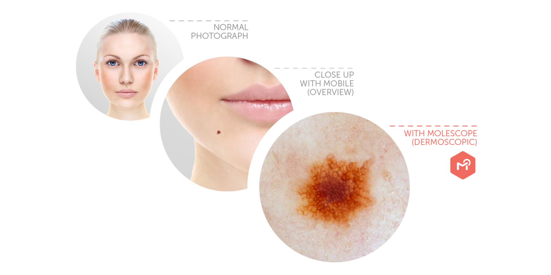
They say a picture’s worth a thousand words. For conditions like skin cancer, where the diagnosis heavily relies upon visual characteristics, capturing and documenting dermatoscopic images should be second nature. Right?
They say a picture’s worth a thousand words. For conditions like skin cancer, where the diagnosis heavily relies upon visual characteristics, capturing and documenting dermatoscopic images should be second nature. Right?
In a study of patients with a high risk of developing melanoma, as many as 50% of malignant cases were only detected with the support of a follow up digital dermatoscopy examination by their care provider.[1] This should come as no surprise, considering how challenging it can be to notice subtle evolutions to the skin over time.
Yet, studies show that less than 43% of GPs use a dermatoscope, and this isn’t even accounting for professionals that use a manual dermatoscope (those without image-capturing capabilities).[2] With over two thirds of Australians being diagnosed with a form of skin cancer in their lifetime, healthcare providers should be equipped with the tools to support the early detection of skin cancer for best possible care outcomes.[3]
Enter the Digital Dermatoscope
A digital dermatoscope is a high-powered camera used to take magnified images of the skin through the use of specialised lighting. This device allows medical professionals to have an enhanced view of a lesion to notice abnormal structures within the skin’s surface that may otherwise be missed.
When examining a patient’s skin, acquiring a baseline image could be the difference between detecting a cancerous evolution in a few months versus years. With the snap of a camera, medical professionals gather vital details about the lesion’s history and have access to a greater depth of information, allowing them to:
- Easily compare skin images: baseline and follow up photos simplify ongoing monitoring and treatment
- Save time: no need to dedicate your valuable time writing down lesion characteristics when you can just snap a picture
- Enhance patient outcomes: increased detection accuracy rates reduce the number of excised benign lesions, saving the patient and physician valuable time, costs, and undue stress
Perfecting Dermatoscopy Workflows
When used effectively, the digital dermatoscope possesses unparalleled potential to support GPs in providing their care to skin cancer patients. Once adopted, what can this look like in daily workflows? With dermatology software playing an increasingly larger role in daily practice, platforms are beginning to offer tools that specifically cater to a faster, easier dermatoscopy imaging experience.
- Immediately Sync Images
One of the biggest reservations people have when taking images is how to properly organise them. The hassle of manual uploads and back ups prevents users from truly taking advantage of the benefits dermatoscopy has to offer. Instead of wasting valuable time, medical systems such as DermEngine are offering secure connected apps which let you capture and sync images directly to the patient’s profile. Eliminating the heavy lifting for documentation processes, GPs can focus on the case itself rather than worrying about ensuring the images make it to the right report.
- Compare Auto-Rotated Images
Perhaps not something typically thought of as a challenge, how well can you compare lesion images taken from drastically different angles? As you’ll find with any task that requires you to spot the difference, having one image “upside down” makes things measurably more difficult. That’s where Image Registration comes in- a simple tap aligns the two dermatoscopic images for easier side-by-side comparison.
Much like documenting the image, it comes down to how much time you’re willing to dedicate to the process. What may seem like a small task at the time can add up to a frustrating experience surprisingly quickly. Once you’ve added Image Registration to your daily routine, it becomes hard to remember how you ever compared images without it.
- Access A Library Of Related Images
When providing a clinical diagnosis, it’s imperative that the medical professional is confident and informed in their professional decision. One tool designed to support this need is DermEngine’s Visual Search. By submitting a dermatoscopic image to the system, Visual Search provides 20 of its most visually similar images from a database of thousands of pre-labelled images in a matter of seconds. Acting as an encyclopedia for skin cancer, the GP can review the images, gain new insights, and learn about the history of these visually similar cases to make a confident, evidence-based decision.
Dermatoscopy Made Accessible
When given the opportunity, the dermatoscope makes a powerful ally. As skin cancer rates continue to rise in Australia, it’s important that healthcare providers are equipped with the tools to offer their services to patients. For GPs who are ready to get started with dermatoscopy imaging, enter the discount code TMR15 at checkout to receive 15% off MoleScope II- a mobile dermatoscope specifically designed for primary care providers.
[1] https://www.ncbi.nlm.nih.gov/pmc/articles/PMC6092075/
[2] https://www.ncbi.nlm.nih.gov/pmc/articles/PMC6502297/
[3] https://www.cancer.org.au/about-cancer/types-of-cancer/skin-cancer.html


