Skin cancers diagnosed by automated dermatologists? This may become the norm sooner than you might imagine
Skin cancers diagnosed by automated dermatologists? This may become the norm sooner than you might imagine
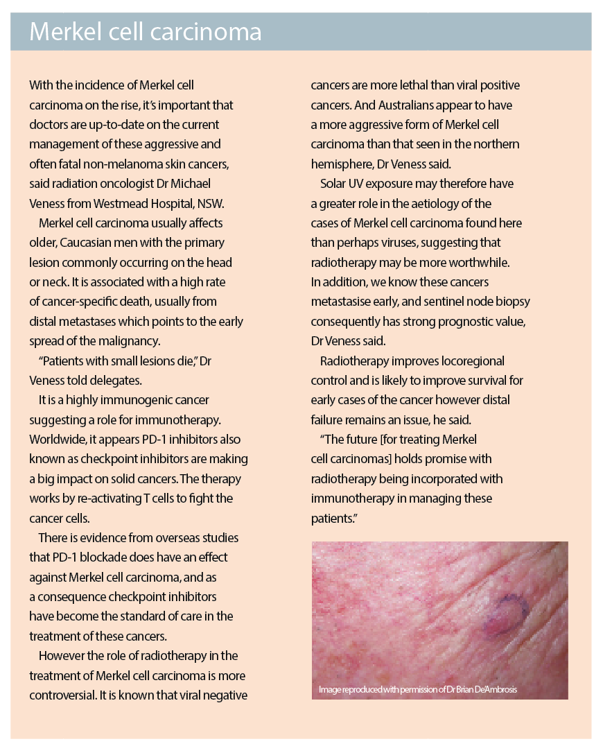
MELANOMA
The reality of melanomas being identified macroscopically by computers rather than humans is fast approaching, US expert Dr Ashfaq Marghoob, director of the Memorial Sloan Kettering’s Regional Skin Cancer Clinic in New York told delegates.
Determining which pigmented lesions should be excised and sent for biopsy has always been one of the great challenges in clinical practice, he said. And we are now very close to have technological detection of melanoma supercede, in both sensitivity and specificity the current methods of detection that have always relied on human interpretation.
The first algorithm to help doctors distinguish melanomas from benign naevi was the ABCDE method of assessment, looking at asymmetry, border, colour, diameter and finally evolution that took into account any recent change. In specialised clinics this method has been found to have a sensitivity of 70% and a specificity of 75%, meaning that one melanoma was diagnosed for every 12-15 lesions excised.
Technology was first utilised in the world of melanoma detection back in the late 70s, with the incorporation of total body photography into clinical practice. This was particularly useful for patients with multiple naevi.
Research, published in the late 90s showed the use of total body photography resulted in fewer naevi being removed but not at the expense of thicker melanomas being diagnosed, meaning that sensitivity was not sacrificed when specificity was improved, Dr Marghoob said. It was estimated that this technology had reduced the number needed to treat down to just 10.
Specificity was further enhanced by the development of dermoscopy. While still relying on human interpretation, the addition of dermoscopic diagnostic criteria with photography in specialised clinics further reduced the number needed to treat to 4-7 according to research.
So, can computers do better than clinicians in detecting melanomas?
According to coding experts, everything is an algorithm, Dr Marghoob said, so theoretically computers could be programmed to recognise globules and networks as detected by the human eye via dermoscopy. In fact, when charged with the task, IBM programmers developed their first algorithm within a week. Using diagnostic dermascopic features, the algorithm not only detected melanoma but could also identify BCCs and SCCs.
Probably the most significant evidence that the human brain will soon be replaced by a computer was research published in the journal, Nature earlier this year. The study, from Stanford University researchers, showed how an algorithm was able to detect skin cancer with accuracy equivalent to expert clinicians. They had assessed the sensitivity and specificity of skin cancer identification from a databank of over 130,000 images (that included about 750 melanomas), and found the algorithm was at least as accurate as the 21 dermatologists who were also asked to assess the images.
What’s more, the ability of the program to ‘learn’ from its mistakes, to analyse lesions that had been misdiagnosed and discover new features, that doctors were not aware of, that could help distinguish benign from malignant lesions suggested it was only a matter of time until the ‘automated dermatologist’ surpassed the clinician in judging which lesions needed excision and which were safe to simply watch, Dr Marghoob said.
“It’s not so much artificial intelligence as augmented intelligence,” he concluded.
ALOPECIA
New research suggests environmental factors may contribute to the development of a particular variant of female baldness, dermatologists at the conference were told.
Frontal fibrosing alopecia is a variant of alopecia that appears to be increasingly common among post-menopausal women, said dermatologist Dr Leona Yip from the Australian Catholic University, ACT.
It is characterised by a symmetrical, progressive recession of the frontal hairline of less than one centimetre a year. Eyebrow loss is common and more aggressive cases may be associated with papules at the hairline.
Very little is known about the aetiology of this particular type of alopecia, Dr Yip said. However menopause is known to be a trigger and it has therefore been thought that sex steroids may have a role. Consequently, finasteride and dutasteride have been trialled in some small studies for the treatment of this condition. While there have been no good randomised controlled trials of these treatments, their effectiveness, even in the smaller studies, appears limited with only 30% of women, who have frontal fibrosing alopecia alone showing any improvement.
Of increasing interest is the fact that the vast majority of cases occur in fair-skinned patients suggesting perhaps environmental triggers such as sun exposure or photosensitivity might be involved.
One theory currently being investigated is that frontal fibrosing alopecia is a response to the repetitive exposure to topical allergens. In particular, sunscreens or components of certain sunscreens may be responsible. There is some evidence that shows the prevalence of frontal fibrosing alopecia is higher among women who use daily sunscreen on their face, often as part of their foundation, daily.
It is likely that further patch testing of sunscreens in high-risk individuals will provide more evidence on this association, Dr Yip said.